

-
ProductsRealizing the Concept of "Practitioner of Precision Medicine"
-
SolutionOne-stop Space Biology Solution for Multi-application Scenarios
-
Technical ServicesPerfect multi-omics (DNA\RNA \protein) detection platform
-
About UsBeijing Fino Weikang Biotechnology Co., Ltd.
-
Consulting Dynamics
17
2023
-
11
High-definition panoramic tissue imaging and AI quantitative analysis technology training course ended successfully!
From November 9 to 10, the "High Definition Panoramic Tissue Imaging and AI Quantitative Analysis Technology Training Course" sponsored by the Institute of Brain Science of Fudan University and co-sponsored by Shanghai Jiangwen and various product technology companies was successfully held in Fudan Brain Institute. Major players in various fields gathered together to launch a deep ideological collision.
The following article comes from Jiang Wen scientific research instruments, the author of Shanghai Jiang Wen

From November 9 to 10, the "Training Course on High Definition Panoramic Tissue Imaging and AI Quantitative Analysis Technology" sponsored by the Institute of Brain Science of Fudan University and co-sponsored by Shanghai Jiangwen and various product technology companies was successfully held in Fudan Brain Institute. Major players in various fields gathered together to launch a deep ideological collision.
This training course provides a two-way communication channel between participants and industry experts through guest reports, theoretical lectures and practical operations.
HD Panoramic Tissue Imaging and AI Quantitative Analysis Technology Training CourseOpening Ceremony
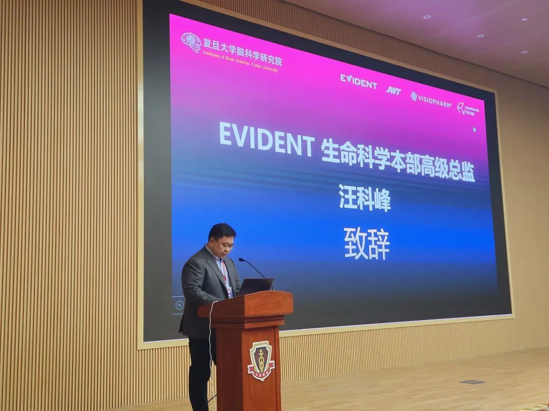
The opening ceremony of this conference was appointed by Jiang Democratic from the Institute of Brain Science of Fudan University and Wang Kefeng, senior director of the EVIDENT Life Sciences Department, extended a warm welcome to all the guests and expressed the center's gratitude to people from all walks of life who have long paid attention to and supported the development of scientific research and technology. I wish this event a smooth development. The conference will write reports on brain diseases, tumors, immunity, nerves and other related topics.
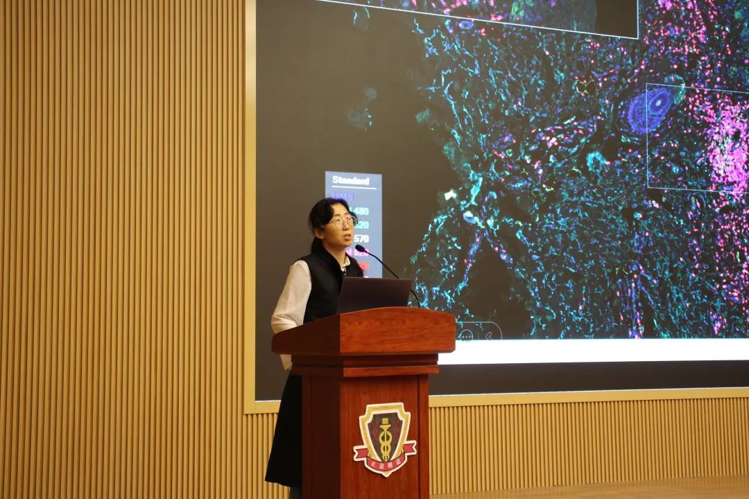
The meeting invited experts from various fields to give reports, share the latest research results, explore scientific problems and exchange innovative opinions.
The conference organizes imaging and analysis techniques around the panorama, from research applications to presentation of technical details.
《Neuroregulation at the Whole Brain Scale: From Anatomy to Function》

Professor Li Wensheng
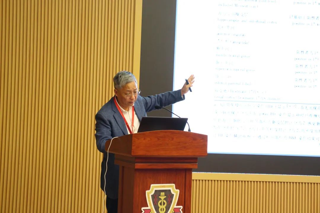
Construction of Fudan Brain Bank and Pathomorphological Analysis of Large Samples
Xiong Man Researcher
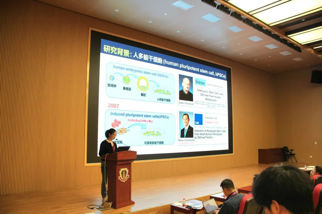
Transplantation Therapy and Disease Simulation of Brain Diseases Using Human Stem Cells
Dr. Huang Ziling
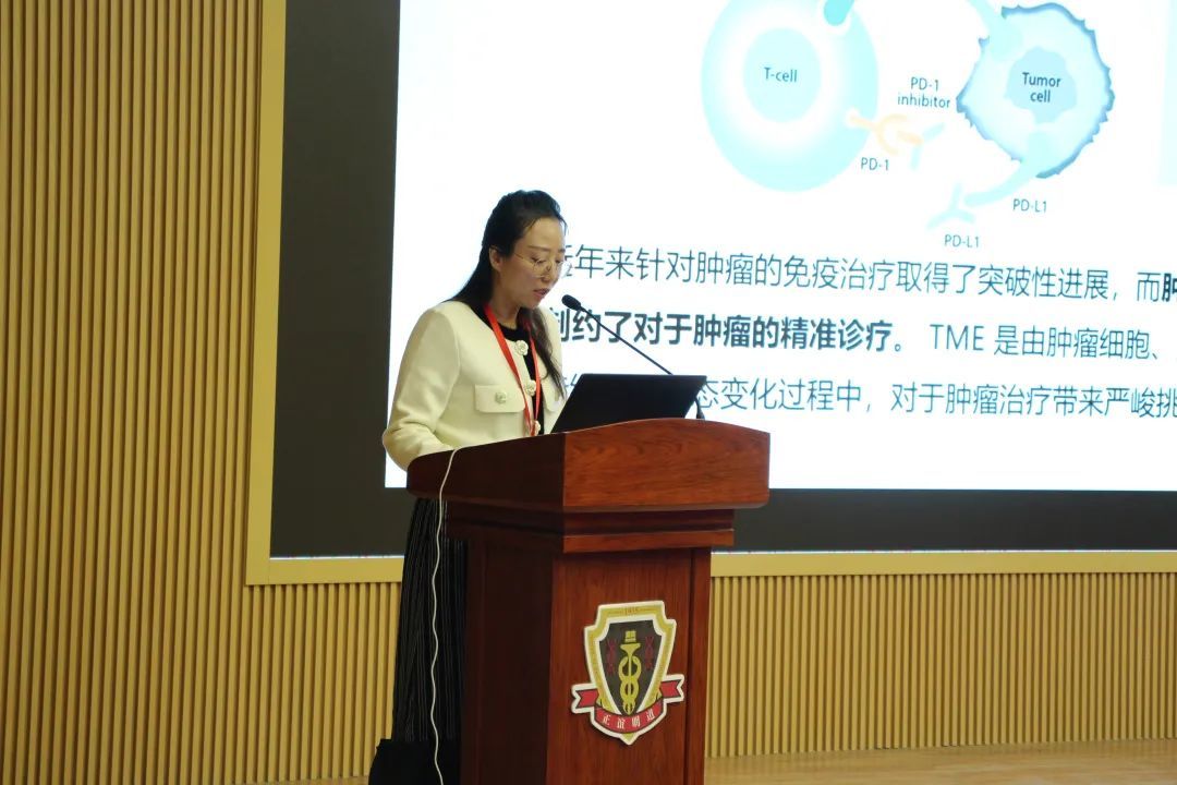
"Research on Tumor Tissue Microenvironment and Exploration of Translational Medicine"
Researcher Min XuThe anatomical position relationship of the brain involved in sleep and wakefulness function is explained from the structure of brain regions, and its precise positioning and functional analysis in the whole brain neural circuit are realized by means of panoramic tissue imaging.
Professor Li WenshengThis paper introduces the development course and the latest progress of the construction of Shanghai brain bank, and displays the histopathological image data of some brain bank samples, which fully reflects the high level of histopathological staining technology and comprehensive imaging analysis technology of Shanghai brain bank workers.
Xiong Man ResearcherThe transplantation therapy and disease simulation of brain diseases using human-derived stem cells were reported, which fully demonstrated the therapeutic potential of human-derived stem cells in brain diseases and the ability to induce neurological diseases as animal models.
Dr. Huang ZilingFrom the perspective of clinical pathology, this paper discusses the significance of tumor microenvironment for clinical tumor diagnosis, and how to accurately guide tumor treatment through panoramic pathology.
Jiang Democratic Ren
Introduction to the Open Platform of Brain Science Research Institute
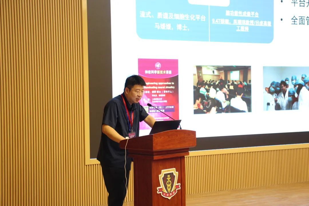
Chen Da
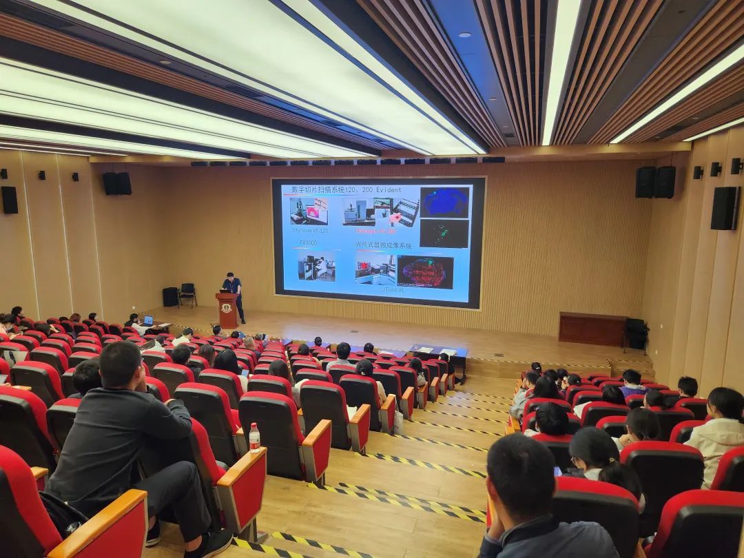
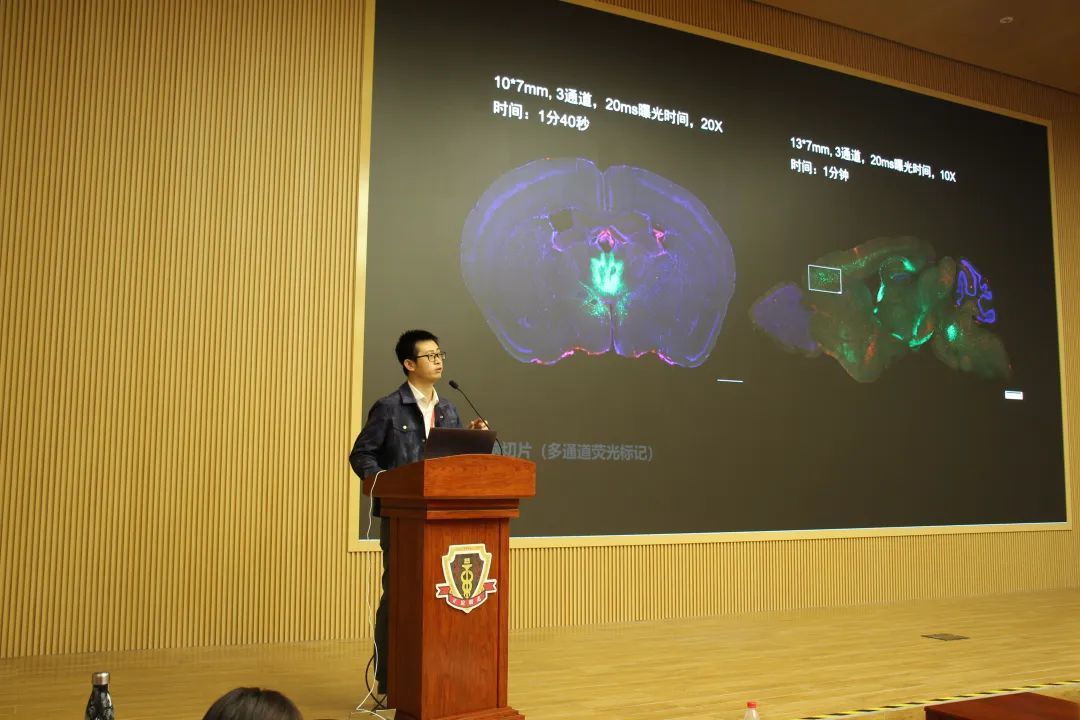
New technology of high-definition panoramic tissue slice scanning
Dr. Pang Shengu
AI Quantitative Digital Pathology Solutions
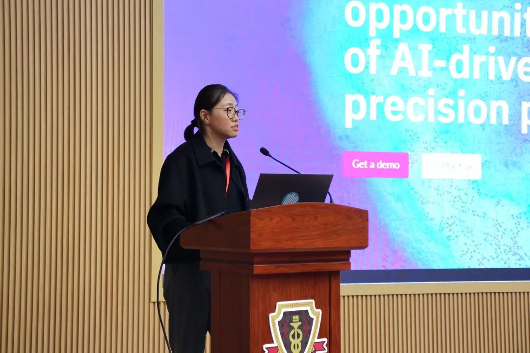
Dr. Li Na
Overall Solution and Application of Tissue Microenvironment Spatial Phenotype Analysis
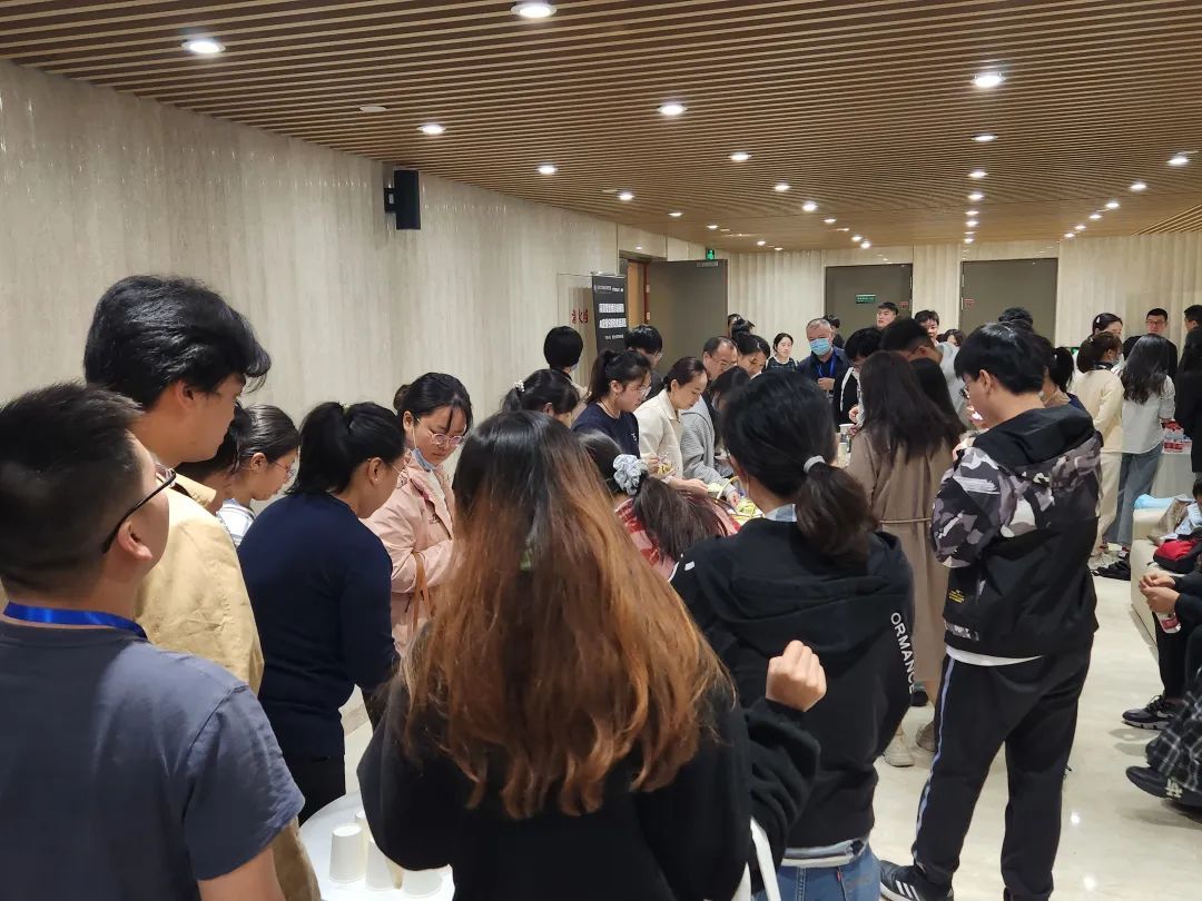
Prepare a fine tea break

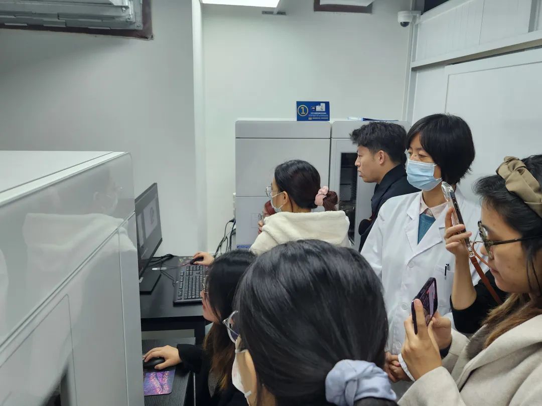
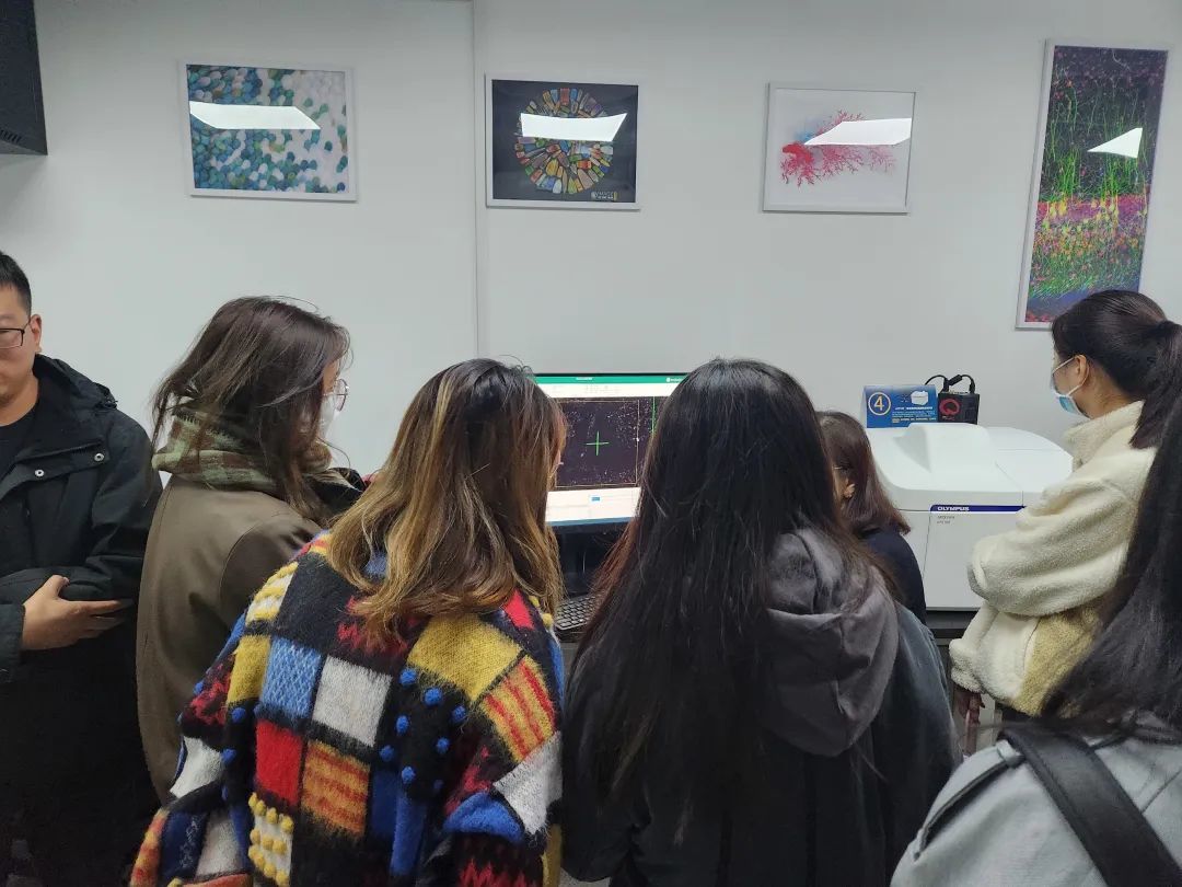
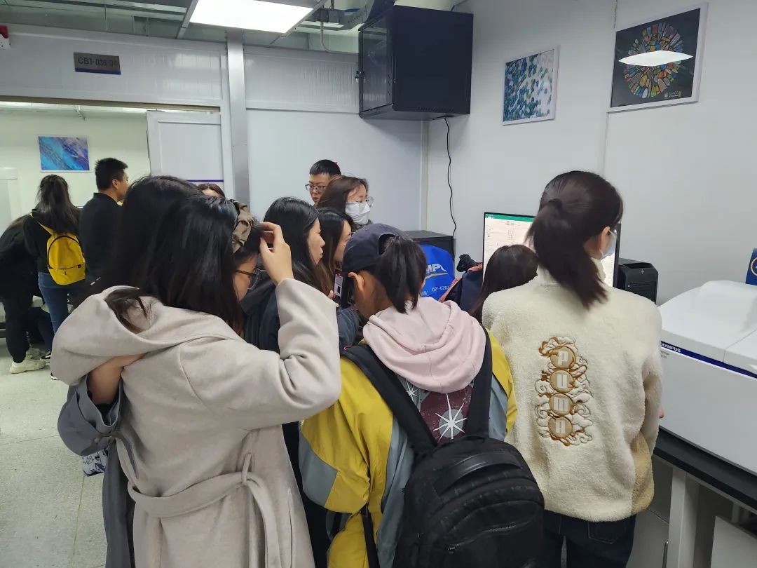
Practical operation demonstration site
On November 10, group practical training was carried out, and participants could carry their own samples to experience the imaging effect. The practical operation includes high-throughput fluorescence digital slice scanner, 3D high-definition side-cutting scanner, tissue microenvironment spatial phenotype analysis system and other cutting-edge technology operation sharing. In this training, professional and technical personnel gave technical explanations to interested participants, covering sample preparation, hardware operation, application sharing, shooting skills, etc., to show the experimental results more intuitively.
This activity covers the joint application of preparation and marking of multi-color fluorescent tissue pathological samples, panoramic high-resolution imaging and AI digital quantitative analysis, combined with the multi-angle field research and sharing of speakers, so that participants have a more comprehensive understanding of panoramic tissue imaging analysis, and this previously complex technology can be more introduced into researchers' experiments.
The successful conclusion of the "High Definition Panoramic Organization Imaging and AI Quantitative Analysis Technology Training Course" cannot be separated from everyone's support. Thank you for your insights and look forward to seeing you again next time!
Key words:
Latest information
Floor 3, Unit 1, Building 15, Jiajie Box Enterprise Exchange, Daxing District, Beijing
Official public number two-dimensional code


