

-
ProductsRealizing the Concept of "Practitioner of Precision Medicine"
-
SolutionOne-stop Space Biology Solution for Multi-application Scenarios
-
Technical ServicesPerfect multi-omics (DNA\RNA \protein) detection platform
-
About UsBeijing Fino Weikang Biotechnology Co., Ltd.
-
Consulting Dynamics
mIF image data analysis service
Introduction to the analytics platform
The rapid development of multicolor immunofluorescence (mIF) technology has made it possible to decode more secrets of the tissue microenvironment in situ at the single-cell level. After obtaining high-quality mIF staining results, the next important question is how to decipher and interpret these results. By introducing Visiopharm's leading image analysis artificial intelligence algorithm, Finovicam has generated the "Pheno whole slide analysis (PWSA) platform" to mine the rich data information of whole slide scan images, so as to deeply interpret the tissue microenvironment and spatial biology information.
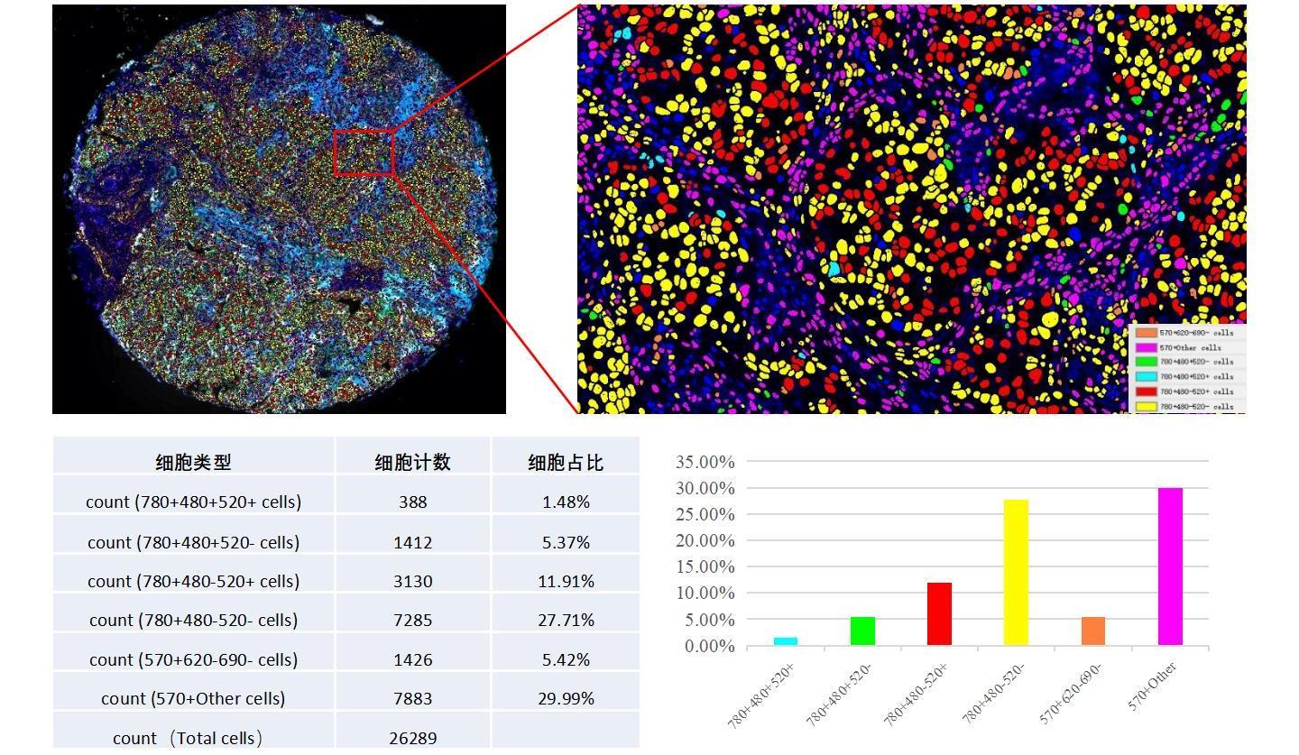
Basic image analysis
Cell protein phenotypic analysis
detection of the number, percentage and density of positive cells (single positive, double positive, multiple positive) of marked proteins

In-depth image analysis

Case 1: Analysis of multiple cell phenotypes
Case 2: Spatial phenotypic and tertiary lymphoid structure analysis of breast cancer samples

Floor 3, Unit 1, Building 15, Jiajie Box Enterprise Exchange, Daxing District, Beijing
Official public number two-dimensional code


