+
  • 试剂盒-1.jpg
  • 试剂盒-2.jpg

Pheno Multicolor Immunofluorescence Assay Kit

Classification:

Reagent

Product accessories:


Contact Us
  • Product Description
  • Operation steps
  • Case Presentation
  • Technical features


    • Up to 7-color labeling on a single tissue section;
    • Signal amplification by 10 to 1,000 times;
    • Reduced usage of antibodies and other reagents;
    • No species restriction for primary antibodies;
    • onservation of samples, protecting precious and rare tissues;
    • Identification of multiple cell phenotypes while preserving spatial localization information;
    • Increased detection combinations of biomarkers, providing special data for multidimensional analysis;
    • The kit includes all reagents required for detection except primary antibodies, offering a one-stop solution without the need to purchase any additional auxiliary reagents./li>


    Technical Principle

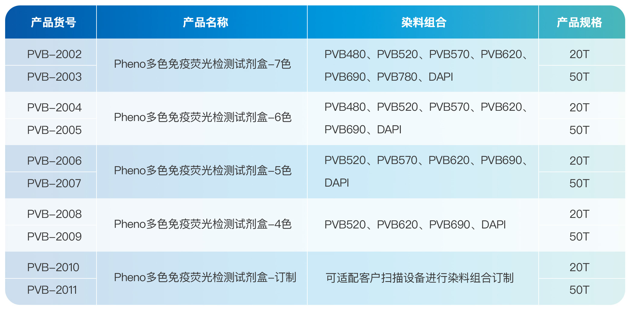
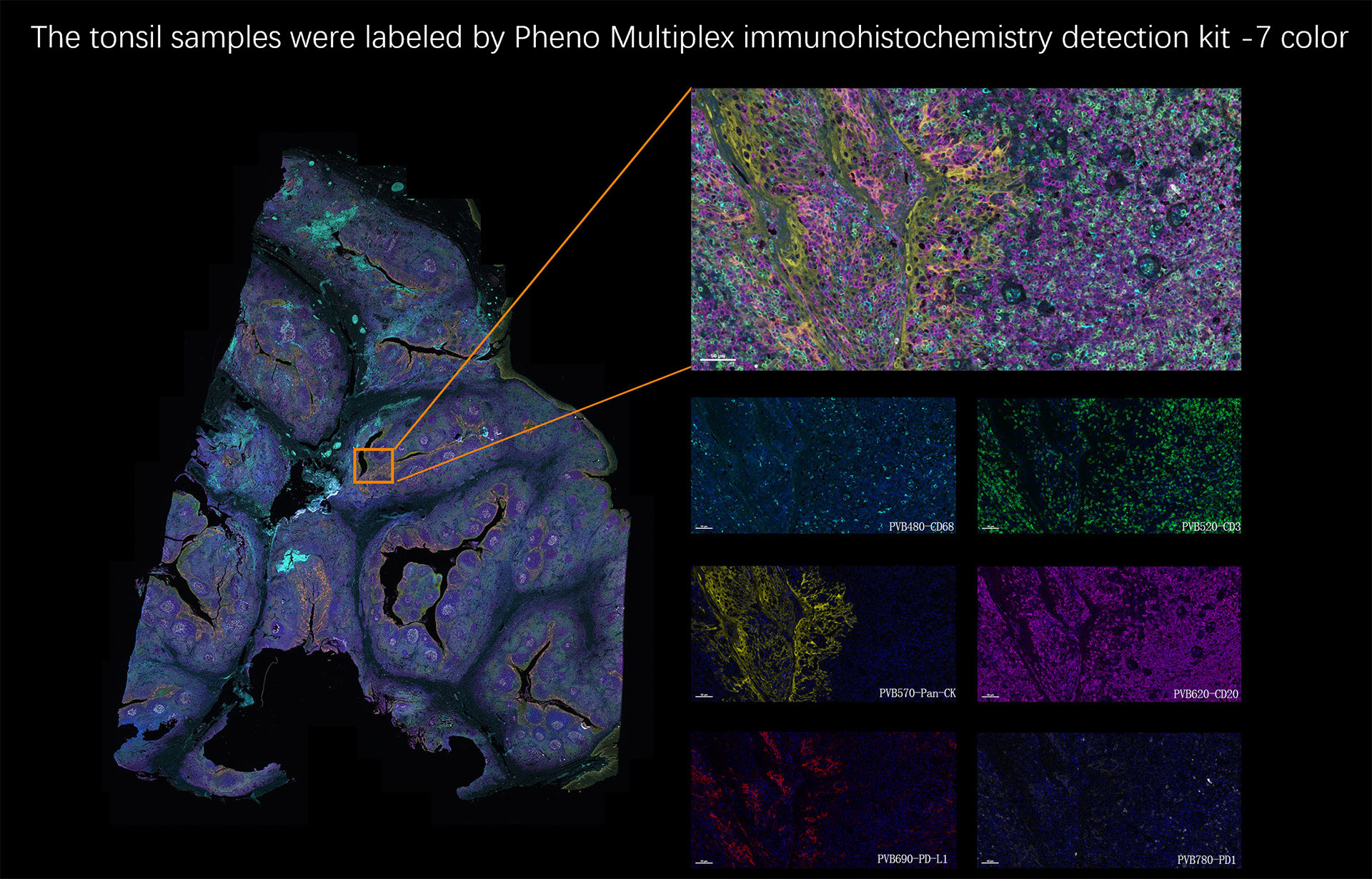
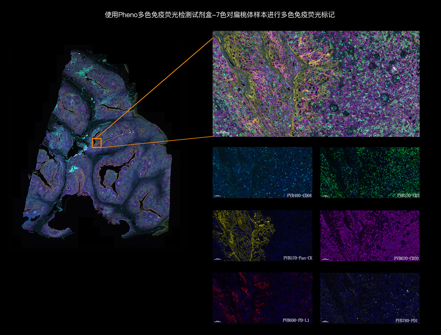
    The Pheno Multiplex Immunofluorescence Detection Kit allows for the simultaneous fluorescent labeling of up to six proteins in FFPE (Formalin-Fixed Paraffin-Embedded) tissue sections. Utilizing Tyramide Signal Amplification (TSA) technology, it can be applied to any type of tissue. This kit enables the study of expression and distribution relationships among multiple proteins at the tissue and cellular levels, making it a powerful tool for in-depth research on tissue microenvironments and the development of biomarkers.


    Operation steps


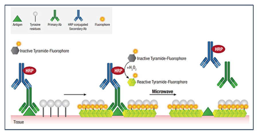
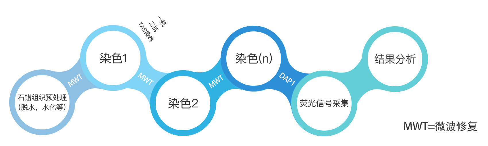
    After sequentially binding the primary antibody, secondary antibody, and TSA fluorophore molecule, the expression of the first protein is successfully labeled. Through microwave repair, the antigen-antibody complexes from the previous round are removed. The fluorophore molecules bound around the antigen are not affected by the microwave or antibody removal and are retained as a fluorescent marker for the antigen-positive cells. In the next round of staining with primary antibody, secondary antibody, and TSA fluorophore, new fluorophore molecules bind around the new target protein. By repeating this fluorescent labeling process multiple times, multiple target proteins can be detected on a single tissue section without the risk of antibody cross-reactivity.


    PhenoVision Bio


    Product Specifications


    PhenoVision Bio


    Case Show


    PhenoVision Bio


  • Operation steps

    After binding the primary anti-secondary anti-TSA fluorescein molecules in turn, the expression of the first protein was successfully marked, and the antigen-antibody complex of the previous round was removed by microwave repair, while the fluorescein molecules bound to the periphery of the antigen were not affected by microwave and antibody removal, thus retaining the fluorescent label of the antigen-positive cells. In the next round of staining of primary anti-secondary anti-TSA fluorescein, new fluorescent molecules are combined around new target proteins; through repeated fluorescent labeling, the detection of multiple target proteins on a tissue section is achieved without the risk of antibody cross-reactivity.


    PhenoVision Bio


    PhenoVision Bio


  • Case Presentation

    PhenoVision Bio


Previous Page

Previous Page